Fig. 4.
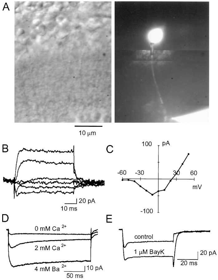
Terminal recordings ofICa obtained with a CsGlu-based internal solution. A, Transmitted light view of a retinal slice (left) in which an RBC has been filled with Lucifer yellow via a recording pipette located on the terminal situated in the most external part of the IPL (right). B, Current records obtained from a different cell under voltage clamp with a patch pipette placed onto the RBC terminal. Depolarizing pulses were applied from a Vh of −70 mV in 20 mV steps.C, Current–voltage relationship for the responses elicited in a terminal recording by depolarizing steps (filled circles; each point is the average of two pulses) (Vh, −70 mV).D, In a different terminal recording, the voltage-gated inward currents were abolished in the absence of external Ca2+, and their magnitude was increased when replacing 2 mm Ca2+ with 4 mm Ba2+. As in the somatic recordings,ICa inactivation was slow. E,ICa elicited by depolarizing steps from a Vh of −70 mV to −10 mV in terminal recording (traces are the average of 24 individual currents; same cell as in A–C) was enhanced by Bay K 8644 (1 μm). Internal solution for A–E was CsGlu.

