Fig. 8.

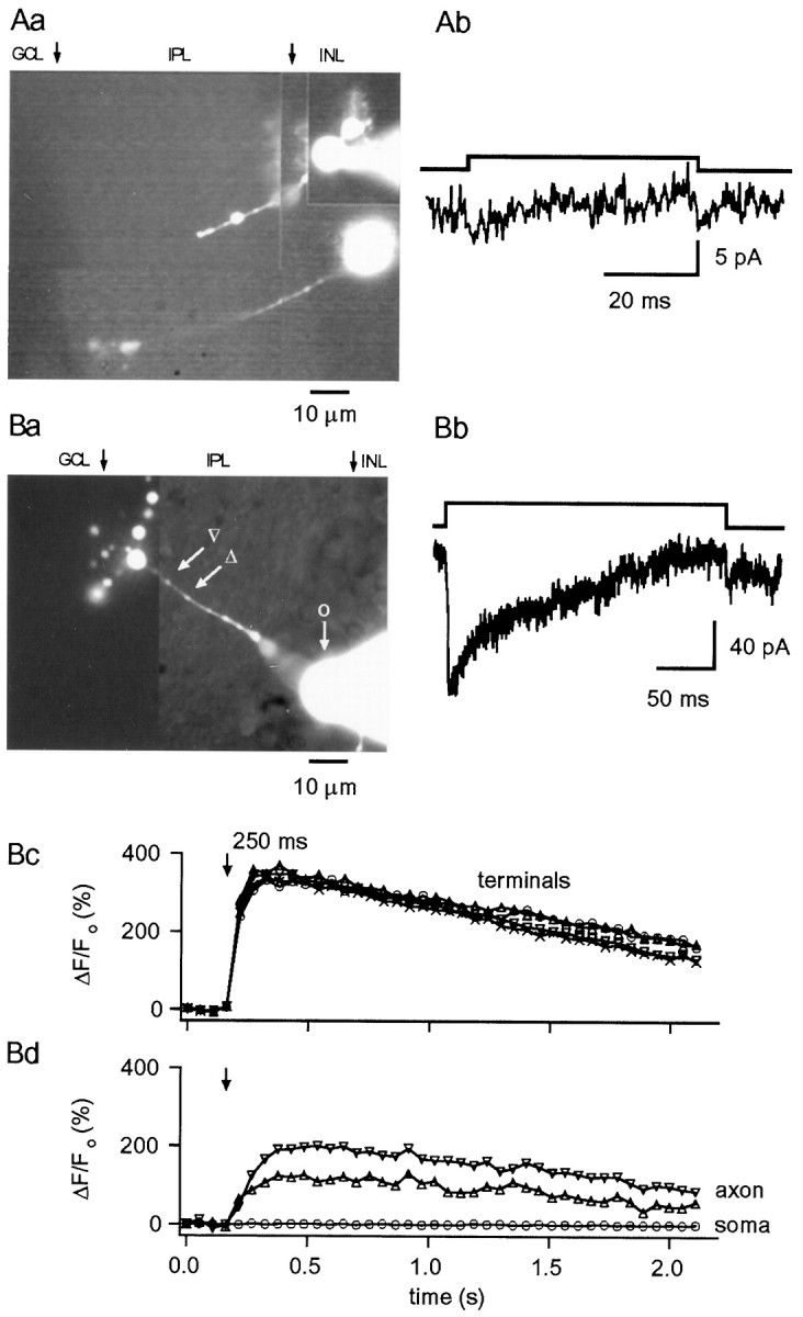
A, Lack ofICa in RBCs lacking terminals.Aa, Photomontage of an area of a retinal slice in which two bipolar cells were recorded; the top cell (with the pipette attached to its soma) lacks part of the axon and the terminal, and depolarizing pulses to −20 mV from a Vh of −70 mV failed to elicit ICa (Ab). Internal solution was CsGlu. B,ICa and [Ca2+]i rises in CBCs.Ba, Photomontage from OG5 fluorescence images showing a CBC with numerous synaptic terminals. Bb, TheICa elicited in this cell (internal solution was CsGlu) by 250 msec depolarizing pulses to −30 mV from a Vh of −70 mV shows pronounced inactivation.Bc, Bd, Depolarization-evoked change in fluorescence were not only observed in the bipolar terminals but also in the enlarged regions of the axon. No changes were detected in the soma. Internal solution was CsGlu (200 μm OG5, no EGTA). Photomontages in Aa and Ba were constructed from images taken at the end of the experiments, as detailed in Materials and Methods. Arrows indicate the approximate boundaries of the different layers identified under transmitted illumination. INL, Inner nuclear layer;IPL, inner plexiform layer; GCL, ganglion cell layer.
