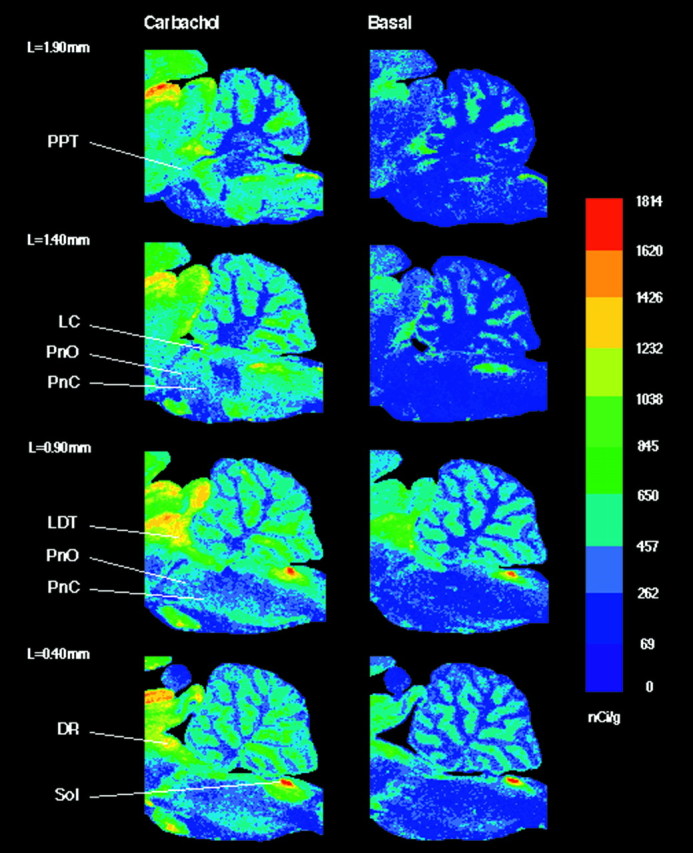
Fig. 3.

Color-coded autoradiograms of sagittal brainstem sections from four different lateralities (L =1.90–0.40 mm). Sections in the left column were treated with 1 mm carbachol, and sections in the right column were treated without agonist. Color bar, Total [35S]GTPγS binding in nanocuries per gram. In the medulla, the nucleus of the solitary tract (Sol) revealed a significant (33.3%) increase in carbachol-stimulated [35S]GTPγS binding compared with basal. Sagittal sections illustrate G-protein activation by carbachol across the rostrocaudal extent of the brainstem.DR, Dorsal raphe nucleus; LC, locus coeruleus; LDT, laterodorsal tegmental nucleus;PnC, nucleus pontis caudalis; PnO, nucleus pontis oralis; PPT, pedunculopontine tegmental nucleus; Sol, nucleus of the solitary tract.
