
Fig. 2.
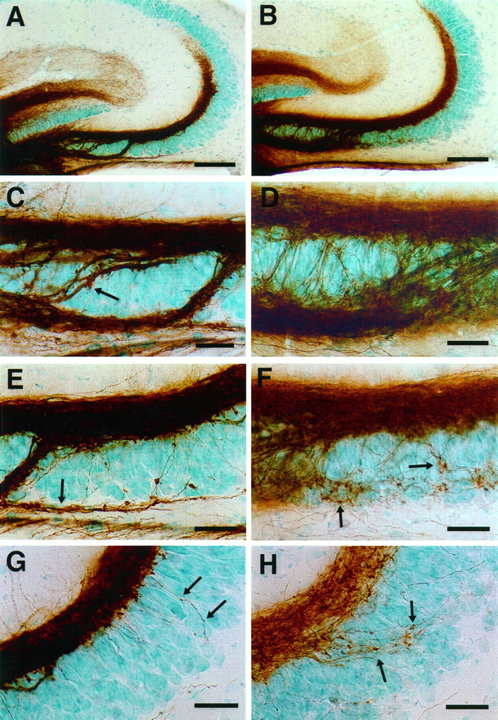
Mossy fiber distribution in 15-d-old control (A, C, E,G) and endo N-injected (B,D, F, H) CF1 mice. The mossy fibers were labeled with DiI with photoconversion to a DAB reaction product (see Materials and Methods). A, B, Low-magnification micrograph showing that the large suprapyramidal mossy fiber bundles are similar images in control (A) and endo N-treated mice (B). C, D, Higher-magnification image of the CA3c subfield. In control mice (C), the intrapyramidal (small arrow) and infrapyramidal (large arrow) mossy fibers are compactly bundled. In endo N-treated mice (D), these bundles are more loosely arranged.E, F, Higher-magnification image of the CA3b subfield. In control mice (E), the infrapyramidal mossy fibers (Figure legend continues) (arrow) run as a fascicle below the pyramidal cell layer. In endo N-treated mice (F), the infrapyramidal mossy fibers are unfasciculated and wander through the pyramidal cell layer, often forming synapse-like structures on the pyramidal cells (arrows). G, H, Higher-magnification image of the CA3a subfield. In control mice (G), fibers arising from the suprapyramidal mossy fibers penetrate into the pyramidal cell layer and display small varicosities. In endo N-treated mice (H), such fibers are more numerous and possess a number of clearly defined synaptic terminals. Scale bars: A, B, 100 μm; C–H, 25 μm.