
Fig. 3.
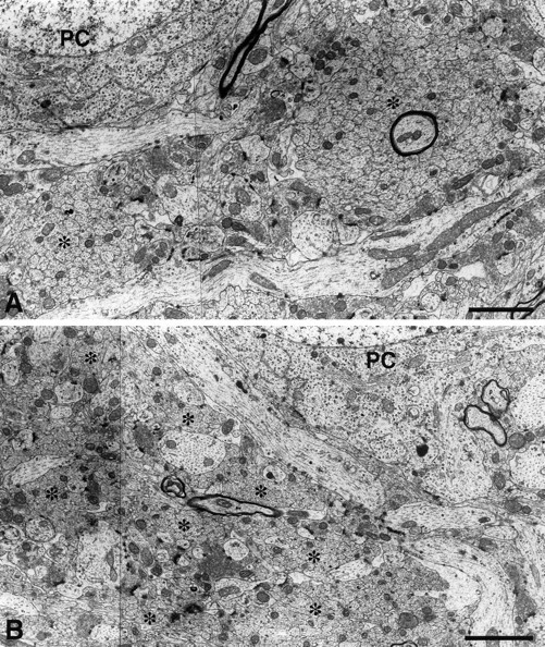
Electron micrograph of infrapyramidal mossy fibers in the CA3c subfield of 15-d-old control (A) and endo N-injected (B) CF1 mice. Sections were cut perpendicular to the mossy fibers. In control mice, unmyelinated mossy fiber axons were tightly fasciculated in large bundles (asterisks), whereas in endo N-treated mice, the fascicles were much smaller and disorganized, with many other elements such as basal dendrites and terminal boutons penetrating into the bundles. Scale bar, 2 μm.