
Fig. 6.
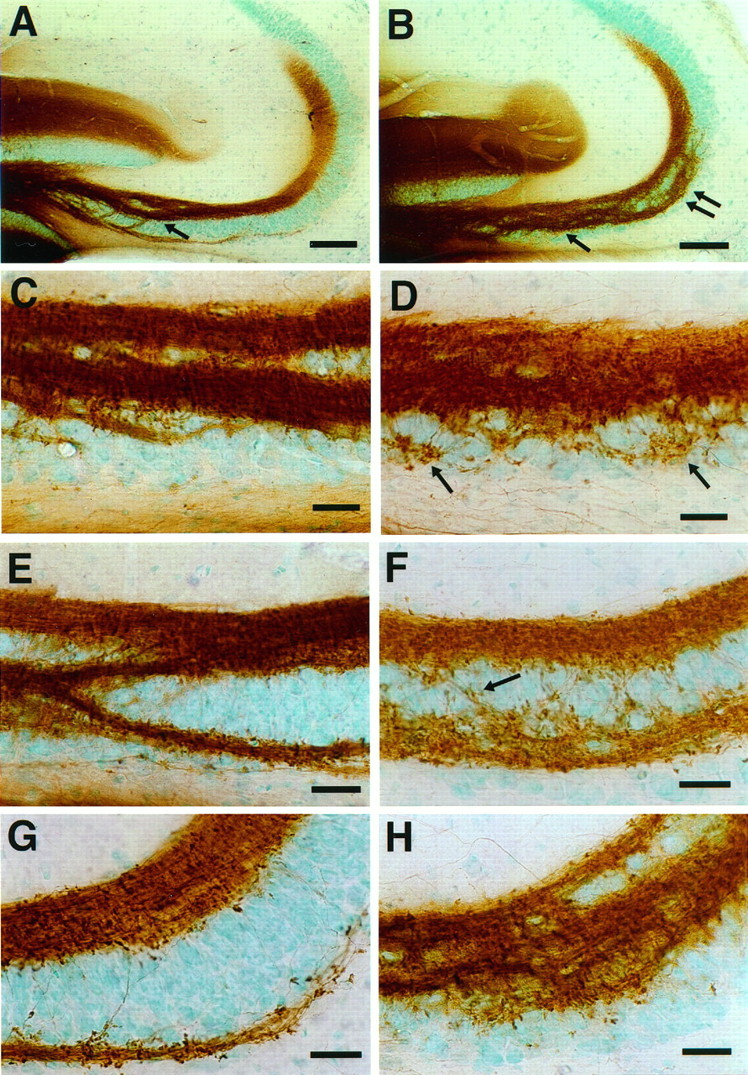
Mossy fiber distribution in 1-month-old 129/SvJ wild-type (A, C,E, G) and NCAM-180-deficient (B, D, F,H) mice. The mossy fibers were labeled with DiI and the fluorescence photoconverted to a DAB reaction product.A, B, Low-magnification micrograph showing that large mossy fiber bundles invade the CA3c pyramidal cell layer in both 129 wild-type and NCAM-mutant mice. However, only in the mutant do these fibers extend into the CA3a subfield (double arrow). C, D, Higher-magnification image of the CA3c subfield. In the mutant, a larger number of fine mossy fibers arise from the intrapyramidal bundle. These ectopic fibers are distributed randomly throughout the lower part of the pyramidal cell layer and make mossy fiber synaptic (Figure legend continues) terminals (arrows). E,F, Higher-magnification image of the CA3b subfield. In wild-type mice (E), intrapyramidal mossy fiber bundles merge with supra- and infrapyramidal mossy fibers. In mutant mice (F), the intrapyramidal mossy fibers track into the pyramidal cell layer with many fine fibers randomly distributed between the supra- and intrapyramidal mossy fibers and the formation of numerous terminal structures. G,H, Higher-magnification image of the CA3a subfield. In the mutant mice (H), a disorganized group of intrapyramidal mossy fibers penetrates into the pyramidal cell layer. Scale bars: A, B, 100 μm;C–H, 25 μm.