Fig. 7.
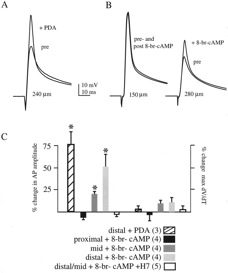
PKA and PKC activation increases the back-propagating action potential amplitude in the distal dendrites.A, Antidromically initiated action potentials before (pre) and after (+PDA) bath application of 10 μm PDA. In this recording 240 μm from the soma, application of PDA lead to a 62% increase in action potential amplitude (from 44 to 71 mV). B, Antidromically initiated action potentials before (pre) and after (post and +8-br-cAMP) bath application of 100 μm8-br-cAMP. Left, In a more proximal recording (150 μm from the soma), in which A-channel density is smaller, action potential amplitude was large to begin with, and 8-br-cAMP did not lead to an increase in amplitude. Right, In this distal recording (280 μm from the soma), in which action potential amplitude is attenuated because of high A-channel density, PKA activation increased the amplitude over 40% (from 33 to 47 mV). C, Summary data. Percent change in action potential amplitude and maximal rate of rise are plotted for five conditions: distal recordings after PDA application, proximal (150 μm) recordings after 8-br-cAMP application, mid (180–200 μm) recordings after 8-br-cAMP application, distal (250–320 μm) recordings after 8-br-cAMP application, and distal and mid recordings after 8-br-cAMP application with 300 μm H7 included in the external solution. Thenumber of cells for each group is inparentheses. Asterisks denote a significant percent increase over the controls (see Materials and Methods).

