
Fig. 6.
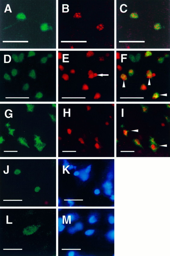
Confocal microscopic images document the localization of caspase-3p20 immunoreactivity and TUNEL in single tissue sections (40 μm) from normal cerebellar hemisphere (postnatal day 5, P5) (A–C) and ischemic striatum (D–F) and parietal cortex (G–I) after 2 hr middle cerebral artery occlusion and 12 hr (D–F) or 24 hr (G–I) reperfusion. Immunoreactivity and TUNEL were visualized with Bodipy fluorescein (A, D, G:green) and Cy3 (B, E, H:red), respectively. Almost every caspase-3p20 immunoreactive cerebellar granule cell at P5 was TUNEL positive. Two representative cells are shown here (C). Many ischemic cells contained both immunoreactive staining (p20) and TUNEL (F, I, arrowheads). Note a typical apoptotic body in a TUNEL-positive striatal cell (E, arrow). After 12 hr of reperfusion caspase-3p20 staining looked as if it was localized within the nucleus of some striatal cells [immunofluorescence staining (J) combined with Hoechst 33342 staining (K) using conventional fluorescence microscopy]. Within cortical cells, however, the pattern of p20 differed at 24 hr (L, p20 immunostaining; M, Hoechst 33342 staining), which may reflect the more rapid rate of ischemic evolution in striatum. Scale bar, 10 μm.