
Fig. 2.
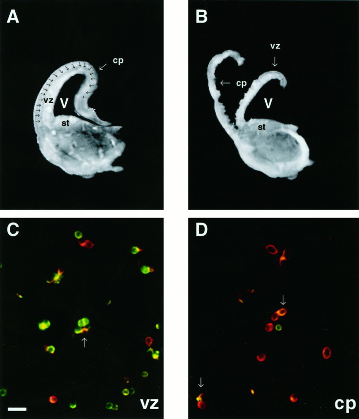
The cortical plate can be microdissected from the ventricular zone in coronal sections of the rat cortex.A, A 350 μm cross section of one hemisphere from an E18 brain. The intermediate zone (iz,arrows) lies between the cortical plate (cp) and the ventricular zone (vz).B, The cross section has been dissected along theiz, resulting in the separation of the cpfrom the vz. Cells in preparations from both regions contain iz cells. C, D, Photomicrographs of cells from each dissociate are double-labeled for nestin (green) and TUJ1 (red).C, The majority of cells in the vz preparation labels with anti-nestin antibody. Other TUJ1+ cells are scattered throughout the field. D, Most cells derived from the cp label with TUJ1, indicating that they are neurons. A minority of cells express nestin only. Arrows inC and D denote newly generated neurons that appear yellow/orange, labeling for both TUJ1 and nestin. st, Striatum; V, ventricle. Scale bar in C, 20 μm.