
Fig. 1.
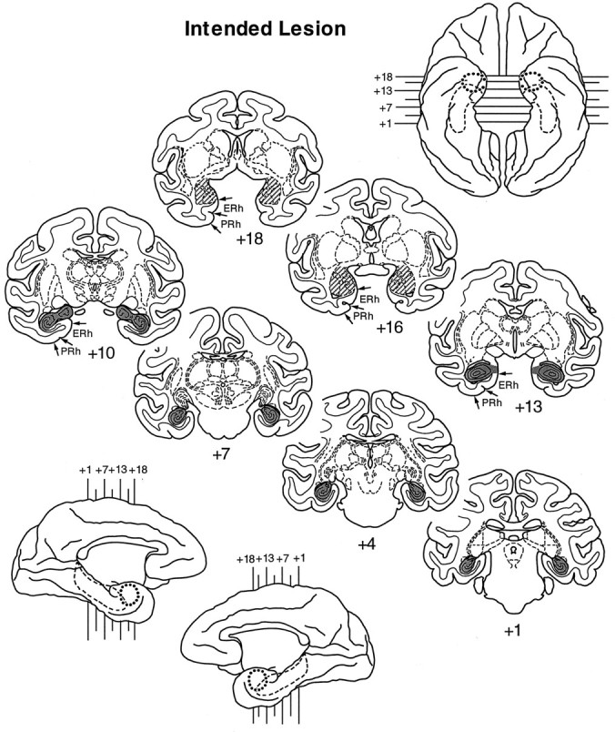
Shaded regions indicate the location and extent of the intended lesion of the amygdala (oblique hatching) and hippocampus (gray) on standard coronal sections. Ventral (top right) and medial (bottom left) views of a standard rhesus monkey brain show the locations of these deep temporal lobe structures: amygdala, dotted line; hippocampus, dashed line. In addition, small arrows mark the boundaries of the entorhinal (ERh) and perirhinal (PRh) cortex, regions we intended to spare, on the coronal sections only (left hemisphere; +18, +16, +13, +10). Thenumerals indicate the distance in millimeters from the interaural plane.