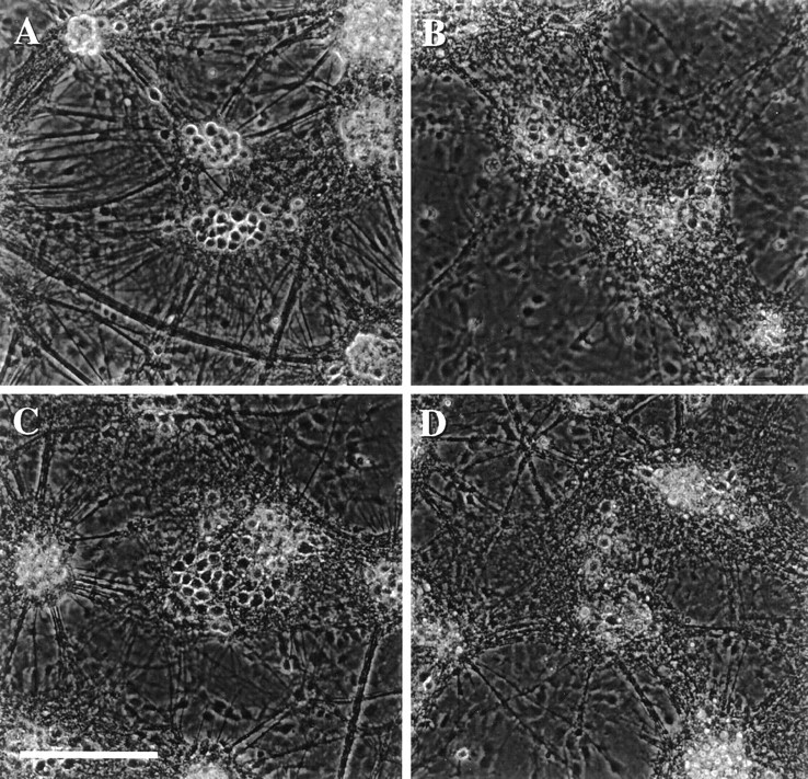
Fig. 1.

Acidic extracellular pH exacerbates slowly triggered AMPA toxicity. Phase-contrast micrographs of mixed cortical and glial cell cultures taken 18 hr after (A) 4 hr exposure to pH 6.6 alone (no change from sham wash controls); (B) 24 hr exposure to 300 μm NMDA at pH 7.4; (C) 4 hr exposure to 300 μm AMPA plus 10 μm MK-801 at pH 7.4 (this exposure, here and subsequently, was terminated by washing out AMPA and adding 10 μm MK-801 plus 30 μm NBQX); (D) 4 hr exposure to 300 μm AMPA plus 10 μm MK-801 at pH 6.6. Scale bar, 200 μm. All data are representative of at least three separate experiments.
