
Fig. 3.
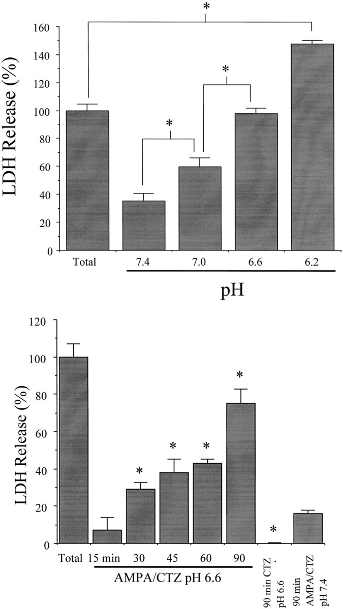
Acidic extracellular pH exacerbates rapidly triggered AMPA toxicity. A, Mixed cultures were exposed for 1.5 hr to 300 μm AMPA plus 100 μmcyclothiazide and 10 μm MK-801 at the indicated extracellular pH (mean ± SEM, n = 4). At pH 6.2 (but not at higher pH), some astrocyte death also occurred, accounting for the total LDH release exceeding 100. *Difference atp < 0.05, as indicated, by one-way ANOVA with Student Newman–Keuls test. B, Time course of rapidly triggered AMPA toxicity at pH 6.6. Neuronal cultures were exposed to 300 μm AMPA plus 100 μm cyclothiazide and 10 μm MK-801 at pH 6.6 for the indicated duration. *Difference at p < 0.05, one-way ANOVA with Student Newman–Keuls test, indicated time AMPA–cyclothiazide (CTZ) at pH 6.6 versus 90 min exposure AMPA–CTZ at pH 7.4 (far right bar). Exposure to cyclothiazide alone at pH 6.6 for 90 min produced no neuronal death (*difference atp < 0.05, 90 min CTZ at pH 6.6 vs 90 min AMPA–CTZ at pH 7.4, Student’s independent t test). LDH release to the bathing medium was measured 24 hr later (mean ± SEM,n = 4) and scaled to the level (100) measured in sister cultures exposed to 300 μm NMDA for 24 hr.