
Fig. 5.
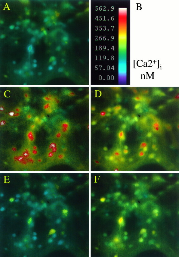
Reducing extracellular pH attenuates the peak [Ca2+]i response to AMPA but delays subsequent normalization. Pseudocolor images, shown as intensity modulated display, of intracellular free calcium in the same field of neurons exposed to 10 μm AMPA at pH 7.4 or 6.6 (24°C).A, Basal [Ca2+]imeasurements in cortical neurons at rest in pH 7.4 buffer.B, Calibration scale.C–D, Peak responses during a 5 sec application of 10 μm AMPA plus 10 μmcyclothiazide and 10 μm MK-801 at pH 7.4 (C) or at pH 6.6 (D).E–F, Corresponding fields asC–D, but measured 15 min after drug washout. Peak [Ca2+]i measurements are reduced by acidic pH, but response recovery is impaired.