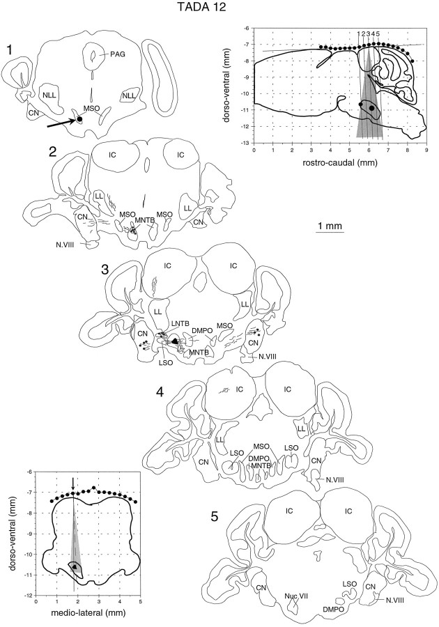
Fig. 2.
Reconstruction of recording sites in one of the bats used in this study. After the stereotactic procedure described bySchuller et al. (1986), the profile of the skull was measured in the sagittal (top right inset) and the horizontal plane (bottom left inset). The profiles were fitted to standard sections as shown in the top right inset. This way the position of the MSO could be predicted with an error of less than ±100 μm. A small HRP injection (arrow) during one of the first penetrations and a large HRP injection (black area in section 3, black dots ininsets) 24 hr before killing the animal were used to confirm the stereotactic calculations and to precisely reconstruct all recording sites. The shaded areas in the two areas give the range of penetrations in this particular animal. The tilted lines labeled 1–5 (top left inset) give the planes of sectioning.

