
Fig. 8.
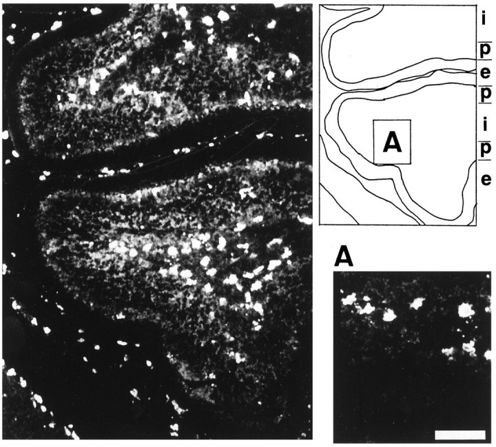
Confocal microscopic image showing the distribution of MRF-1-immunoreactive antigen in P7 rat cerebellum. A section of the P7 rat cerebellum was immunostained with anti-MRF-1 antibody and photographed. Scale bar (shown in A): 50 μm. i, Internal granule layer and/or white matter;p, Purkinje cell layer; e, external granule layer.