
Fig. 9.
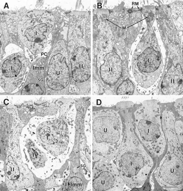
Type I morphologies in the early postnatal mouse utricle. A, B, P4, extrastriolar region fixed in situ. C, P7, striolar region fixed in situ. D, Juxtastriolar region cultured on P1 and fixed 6 d later (P7). On P3–P4, some type I hair cells lacked calyces (Fig. 8C), and others had partial calyces (A, PC) or full calyces (B, I). Some type I cells (B) had the necks that are characteristic of mature type I cells, and others did not (A). The neck is not a response to the calyx, because it was sometimes seen in cultures (D). As the type I cells developed necks, their nuclei tended to move toward the base of the epithelium; compare the adjacent type I cells in D. Complex calyces, which surround more than one type I cell (C) and are common in the mature striola, were first seen on P7. The bulging apical surfaces of the type I cells in D are a secondary trait. In B, the apical reticular meshwork (RM) of a supporting cell is labeled. Other labels are as in Figure 8. Scale bar, 5 μm.