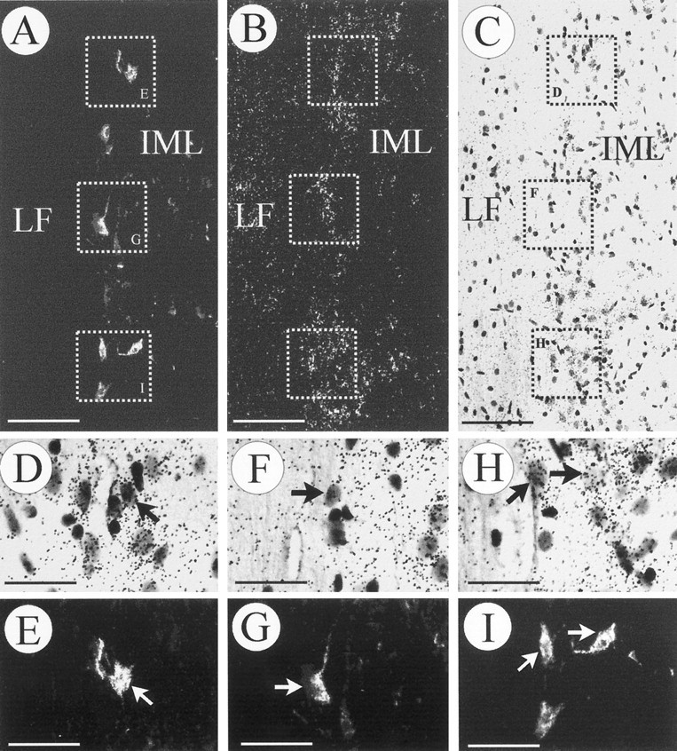
Fig. 4.

Identification of preganglionic sympathetic neurons in the spinal cord. Neurons expressing TrkB mRNA that innervate the adrenal medulla were retrogradely labeled with FG from the adrenal medulla (A, E, G, I) and processed for in situ hybridization (B, C, D, F, H).A–C, An identical longitudinal section (T9–T10) visualized in dark field for the FG label (A), TrkB mRNA (B), and bright field (C). Three areas have been depicted and shown atD/E, F/G, and H/I at higher magnification. Arrows point at FG-labeled cells expressing TrkB mRNA. As expected, a majority of TrkB mRNA-positive neurons, which project to sites other than the adrenal medulla, were not labeled with FG (D/E, F/G, H/I). Scale bars:A–C, 100 μm; D–I, 50 μm.
