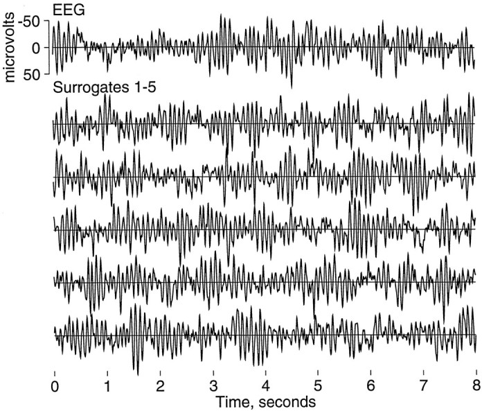
Fig. 2.

EEG and surrogate data. This figure displays a sample 8 sec EEG epoch in the top trace and five surrogate time series generated from it in the tracesbelow. Because the linear structure of the time series is preserved in surrogates, the original time series and its surrogates appear similar by visual inspection. This EEG is representative of the posterior dominant rhythm of α-activity typical of eyes-closed human EEG. Other patterns with more or less α-activity also were observed.
