Fig. 1.
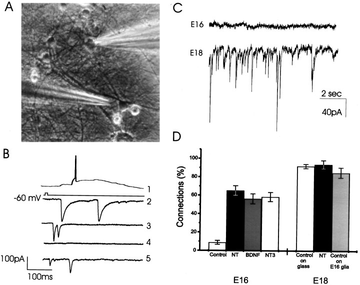
BDNF- and NT-3-induced functional synapses in hippocampal neurons in culture. A, An image of a field in E18 culture. One cell is patch clamped (right shadow), and an adjacent cell is approached with a glutamate-containing pipette (left shadow). Magnification, 40×. B, Trace 1, Illustration of the response of an afferent cell to the glutamate pulse with an action potential. Traces 2-5, Postsynaptic recordings in a single patch-clamped cell after stimulation of four afferent neurons to illustrate the variety of synaptic responses. C, Top trace, Spontaneous activity recorded from a cell taken from E16 and grown in culture for 2 weeks. Bottom trace, Recording from an E18 culture showing extensive EPSCs and IPSCs. D, Two week treatment of the E16 cultures with 20 ng/ml BDNF and/or 20 ng/ml NT-3 caused a marked increase in the levels of functional synaptic connectivity among neurons. In control cultures, only 8.8 ± 2.1% of all tested afferents were connected (n = 52 cells), however, the percentage of connected afferents reached 65.3 ± 5.2% (n = 36 cells), 56.4 ± 5.4% (n = 41 cells), or 58.6 ± 4.9% (n = 38 cells) in BDNF plus NT-3-treated (NT), BDNF-treated, or NT-3-treated cultures, respectively (Fig. 1D). The neurotrophins (NT) did not have a similar effect in the E18 cultures; in both control and NT-treated cultures the majority of cells (>90%, Fig. 1D) were connected. Control E18 neurons also form connections when they are plated at relatively low density on top of monolayer of type I astrocytes (Fig.1D, E18, shaded bar).

