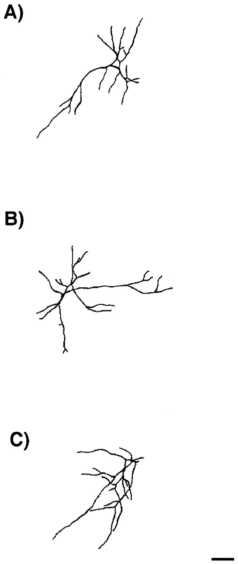
Fig. 8.

Morphology of excitatory E16 neurons in culture. Cells were grown for 14 d in control cultures (A) or in the presence of NT-3 (B) or BDNF (C) from days 11–14. They were fixed and double immunostained for the expression of MAP2ab and GABA. Camera lucida drawings of dendrites and cell bodies were done on MAP2ab-positive/GABA-negative neurons using a 63× objective. Drawings were digitally scanned and processed using Adobe Photoshop. Neurotrophins did not cause a change in dendritic morphology of the excitatory neurons. See Figure 6 for quantitative analysis of morphology. The thickness of the dendrites is represented arbitrarily. Scale bar, 50 μm.
