
Fig. 2.
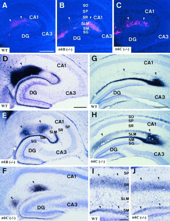
Distribution of entorhinohippocampal and commissural projections in trkB and trkC (−/−)mice. A–C, Distribution of entorhinohippocampal afferents in wild-type (A), trkB (−/−) (B), and trkC (−/−) (C) newborn mice after injections of DiI in the entorhinal cortex. Labeled fibers (arrowheads) are observed running in the stratum lacunosum-moleculare and in the white matter in all three animal groups. Sections are counterstained with bisbenzimide. D–F, Pattern of medial entorhinal projections in wild-type (D), trkB (−/−) (E), and trkC (−/−) (F) P14 mice after iontophoresis of biocytin in the entorhinal cortex. In all three groups, axons innervate the middle tier of the molecular layer and two patches in the stratum moleculare, in the CA2/CA1 and CA1/subiculum interphase (arrowheads). G, H, Distribution of lateral entorhinal projections in the hippocampus of wild-type (G) andtrkC (−/−) (H) P14 mice, illustrating no major topographic differences in the innervation of the outer molecular layer and stratum lacunosum-moleculare in the CA3 region (arrowheads). I, J, Distribution of commissural fibers in the hippocampus of wild-type andtrkC (−/−) P14 mice after injections of biocytin in the contralateral hippocampus. Commissural fibers innervate the stratum radiatum and the inner molecular layer (arrowheads).EC, Entorhinal cortex; DG, dentate gyrus;SG, stratum granulosum; SLM, stratum lacunosum-moleculare; SM, stratum moleculare;SO, stratum oriens; SP, stratum pyramidale; SR, stratum radiatum. Scale bars:A–H, 300 μm; I, J, 100 μm.