
Fig. 5.
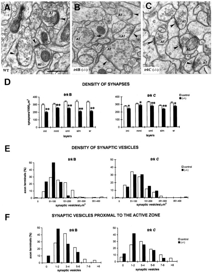
Fine structure of synaptic boutons intrkB (−/−) and trkC (−/−) mice at P13–P14. A–C, Electron micrographs illustrating axon terminals and synaptic contacts (arrowheads) in the stratum radiatum of wild-type, trkB (−/−), andtrkC (−/−) mice. Note decreased densities of synaptic vesicles and reduced thickness of postsynaptic specializations, especially in trkB (−/−)mice. Scale bar, 0.5 μm.D, Density of synaptic contacts in different hippocampal layers in trkB (−/−), trkC (−/−), and littermate controls (mean ± SEM; *p < 0.05; **p < 0.01; Student’s t test).E–F, Histograms showing distributions of density of synaptic vesicles (E) and numbers of synaptic vesicles near the active zone (F) in axon terminals in the stratum lacunosum-moleculare of trkB (−/−) and trkC (−/−) mice and their littermate controls. Note decreased density of synaptic vesicles intrkB (−/−) but not in trkC (−/−) mice, and reduced numbers of synaptic vesicles clustered near the active zone in both mutant mice. AT, Axon terminal;iml, inner molecular layer; mml, medial molecular layer; oml, outer molecular layer;slm, stratum lacunosum-moleculare; sr, stratum radiatum.