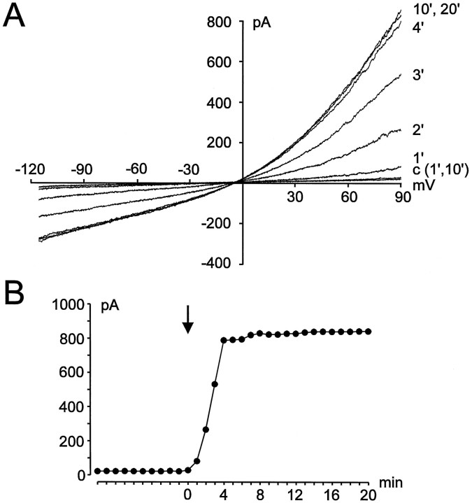
Fig. 3.
Dependence of stretch-activated Cl− currents on intracellular ATP.A, Current recordings during perfusion of the cell with 4 mm ATP. Currents are shown at 1 and 10 min after establishment of the whole-cell configuration before the cell was stretched (c) and at several times (in minutes) after application of the membrane stretch. B, Corresponding amplitudes of the current shown in A(measured at +90 mV) are plotted against time after break-in. Membrane stretch is indicated by the arrow.

