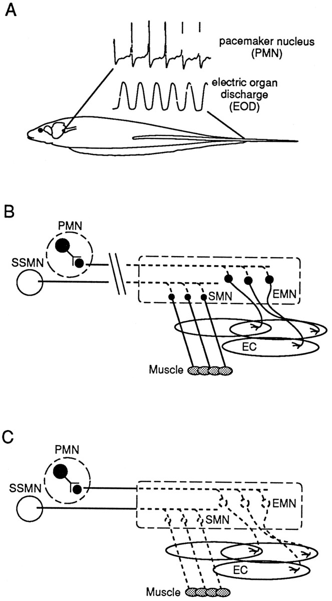
Fig. 1.

Schematic illustration of the electric organ discharge (EOD)-generating circuitry in S. macrurus and its manipulation in the present study.A, Extracellularly recorded field potentials from the medullary pacemaker nucleus (PMN) demonstrate its extremely regular firing frequency. A pulse from the electric organ follows each spike by a few milliseconds. B, Diagram showing the organization of the EOD-generating circuitry and the site of spinal cord transection that removes supraspinal input to both electromotoneurons (EMN) and somatomotoneurons (SMN) and renders the EO and muscle fibers inactive. Axons from the PMN and the summed supraspinal inputs to SMNs (SSMN) distal to the cut degenerate (boxed region), as depicted by the dotted lines. The EMNs and SMNs remain intact (see Results). EC, Electrocytes. C, Diagram showing the elimination of EMNs and SMNs by the removal of a segment of the spinal cord (boxed region), i.e., denervation. Denervation results in the degeneration of motoneuronal axons, leaving the target muscle fibers and electrocytes denervated.
