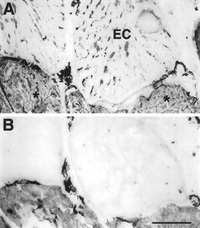
Fig. 12.

Serial sections of a region of a tail 5 weeks after denervation. Electrocytes (EC) are labeled with MF20 (A), but not with 12-101 (B). In contrast, all centrally located muscle fibers (asterisks) reacted positively with both antibodies. The dark label in the extracellular space between electrocytes and muscle fibers corresponds to melanocytes, which always display dark coloration. Scale bar, 400 μm.
