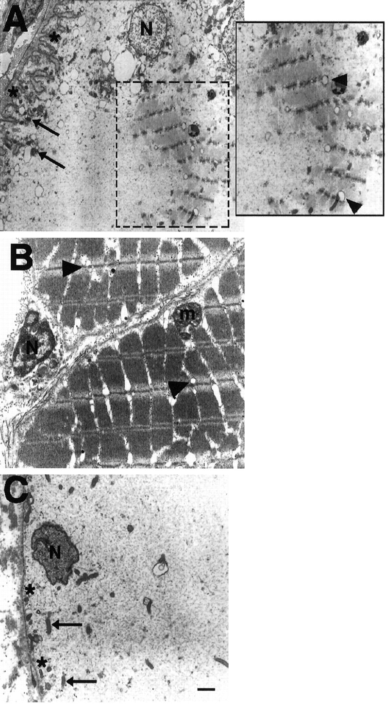
Fig. 13.

Electron micrographs showing regions from an electrocyte 5 weeks after denervation (A), a muscle fiber (B), and an electrocyte (C) from an unoperated control tail. Note the cluster of myofilaments arranged in sarcomeres within the cytoplasm of a denervated electrocyte (A, boxed region). The boxed region is enlarged for clarity of the structures. Note that T-tubules are evident near the myofilaments and aligned with the Z-lines (arrowheads) as they occur in control muscle fibers. The membrane (asterisks) of denervated electrocytes also had many convolutions, a structural characteristic not present in the membrane of electrocytes from unoperated fish. Electrocytes from control fish tails are devoid of sarcomeres. Mitochondria (arrows) and nuclei (N) are located peripherally in electrocytes from both denervated and control tails. Muscle fibers also contain many mitochondria (m) in the cell periphery. Scale bar, 1 μm.
