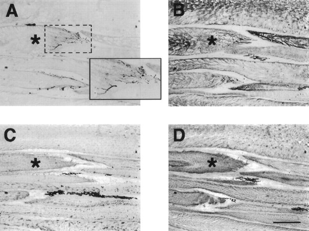
Fig. 6.
Serial longitudinal sections (12-μm-thick) ofS. macrurus tail after 5 weeks of spinal transection show the presence of axons innervating the posterior surface of electrocytes with 3A10 immunolabel (A). The region within the dotted box is enlarged (solid box) to view the labeled axons more clearly. Expression of keratin in all electrocytes was revealed with AE1 immunoreactivity (B). Electrocytes of 5 week transected fish also expressed MHC and tropomyosin, as seen by the positive labeling with antibodies MF20 (C) and CH1 (D). Note the patchy distribution of the label with MF20 and CH1. The asterisk denotes the same electrocyte in all four serial sections. The dark labelin the extracellular space between electrocytes in C andD corresponds to melanocytes, which always display dark coloration. Scale bar, 500 μm.

