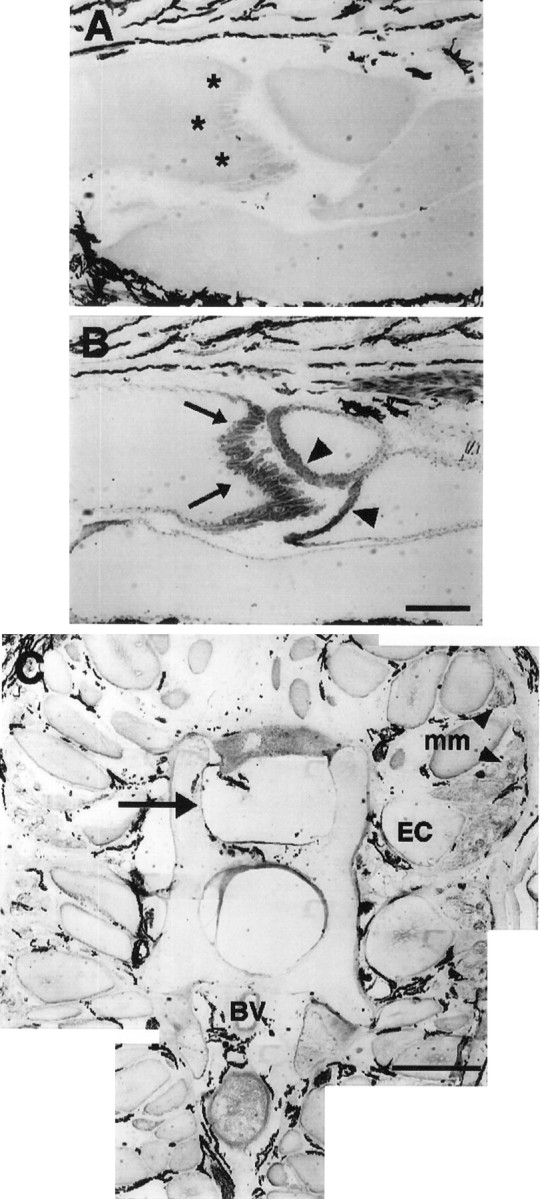
Fig. 7.

Absence of neural tissue within the region of a tail 2 weeks after denervation. The posterior surface of electrocytes (asterisks) is devoid of innervating axons, as shown by the absence of 3A10 immunolabel (A). Immunolabel of acetylcholine receptors with antibody 88b (B) is revealed in both posterior (arrows) and anterior (arrowheads) surfaces of electrocytes. Melanocytes in the extracellular space around electrocytes always display dark coloration. C, Part of a cross section (12-μm- thick) of a 2 week denervated tail near the caudal end of the anal fin and immunoreacted with 3A10. There is no spinal cord within the vertebral column (arrow) or spinal nerves adjacent to vertebrae (compare with Fig. 4C). Dorsal is up; ventral is down. BV, Blood vessel;EC, electrocyte; mm, muscle fibers (arrowheads in C). Scale bars:A, B, 1 mm; C, 200 μm.
