
Fig. 4.
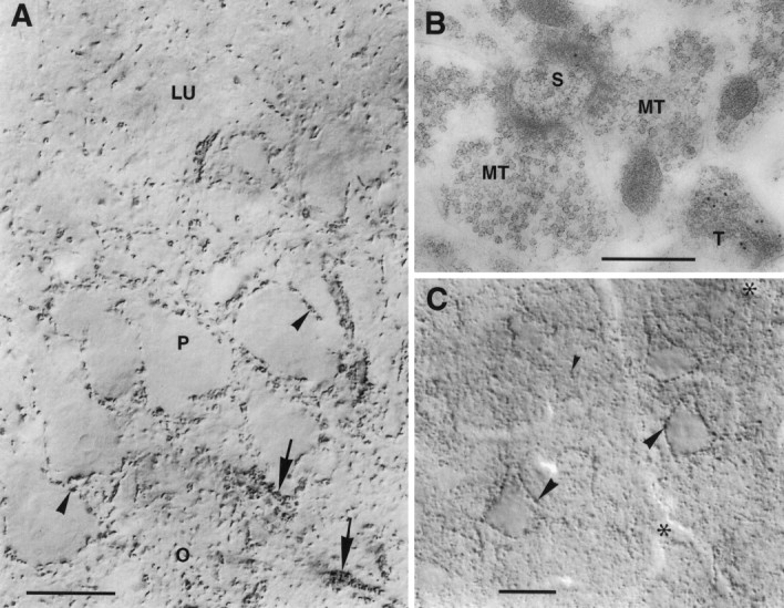
VGAT in hippocampus CA3 and neocortex.A, CA3. Nerve endings immunoperoxidase-stained for VGAT are seen to outline unstained pyramidal cell bodies (arrowheads) and the initial parts of their axons (arrows) or are spread in the neuropil. Mossy fiber terminals in the stratum lucidum (LU) are not visualized. P, O, Strata pyramidale and oriens, respectively. B, CA3, stratum lucidum. Post-embedding immunogold labeling shows VGAT in a terminal with pleomorphic vesicles (T) but not in the large mossy fiber terminals (MT) making asymmetric synapses on a spine (S). C, Limb area of parietal cortex, layer 5. Strongly immunoreactive (peroxidase with Triton) nerve terminals are concentrated along pyramidal cell perikarya (large arrowheads) and are spread in the neuropil (small arrowheads). On the apical parts of the three large pyramidal cells shown, VGAT-containing boutons on the back or front of the cells can be seen en face (in part slightly out of focus).Asterisks, Blood vessels. Scale bars: A, 20 μm; B, 0.5 μm; C, 25 μm.