
Fig. 5.
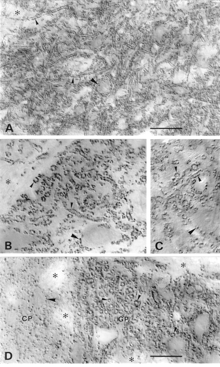
VGAT in basal ganglia and substantia nigra. A–C, Substantia nigra pars reticulata.D, Border between caudatoputamen (CP) and globus pallidus (GP). A,D, N2 antibody with Triton. B, N2 antibody without Triton. C, C1 antibody with Triton. VGAT-containing small nerve endings coat dendrites (small arrowheads) very densely and perikarya (large arrowheads) less completely. The staining pattern in substantia nigra and globus pallidus is typical of the GABAergic afferents from caudatoputamen. Antibodies to the N terminus (A, B, D) and C terminus (C) of VGAT show the same localization with (A, C, D) and without (B) Triton. Asterisks, Bundles of unstained nerve fibers. Scale bars: A, 50 μm;B–D, 20 μm. .