
Fig. 3.
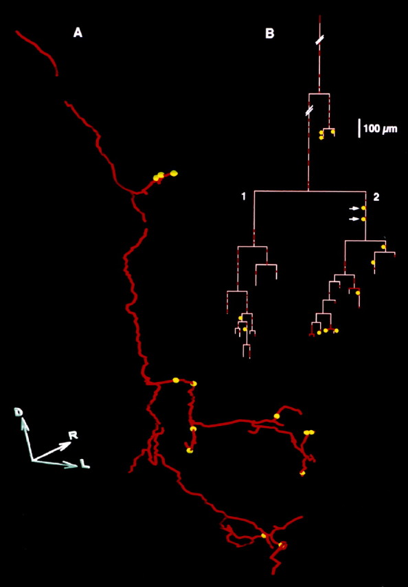
A, Planar projection of the three-dimensional reconstruction of a Ib collateral (red) with thirteen presumed GABA-immunoreactive contacts (yellow spheres). D,R, and L indicate, respectively, the dorsal, rostral, and lateral directions. Length of each axis, 100 μm.B, Axogram of this collateral. The redsegments indicate the segments of the fiber that were examined for GABA immunoreactivity presumed contacts in the most superficial 6 μm of the sections. The pink segments indicate the remaining portions, in which GABA-immunoreactive structures were not detectable (see Materials and Methods). Further comments in Results.