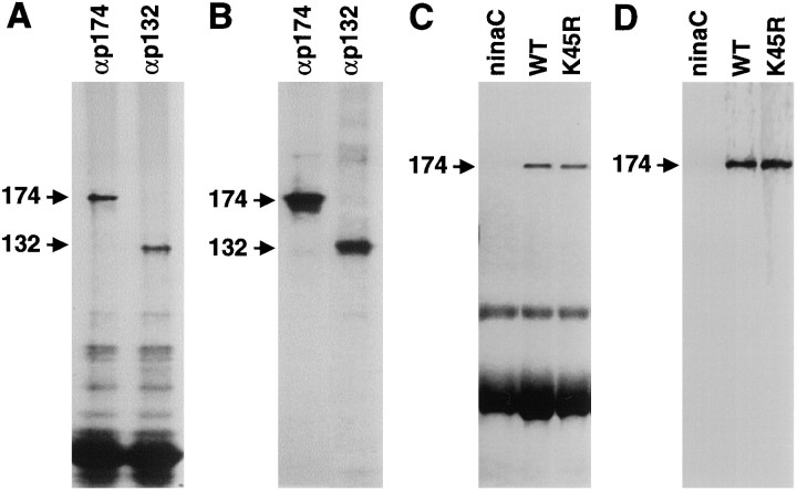
Fig. 1.
NINAC proteins were phosphorylated in vivo. A, Wild-type flies were fed32P-labeled orthophosphate, and the NINAC proteins were immunoprecipitated from head extracts with antibodies to p174 (αp174) or p132 (αp132) and were fractionated by SDS-PAGE. The proteins were detected by staining with Coomassie blue. B, The same gel shown inA is shown exposed to x-ray film. C, Phosphoproteins in wild-type (WT),ninaCP235 (ninaC; null allele), and P[ninaCK45R] (K45R) flies were labeled with [32P]orthophosphate, fractionated by SDS-PAGE, and detected by staining with Coomassie blue. D, The same gel shown in C is shown exposed to x-ray film.

