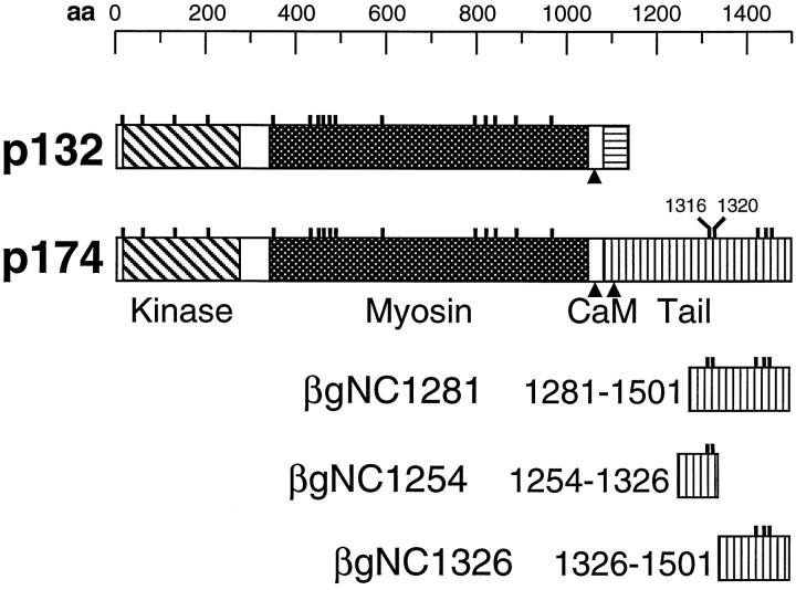
Fig. 2.
Consensus PKC phosphorylation sites are indicated by the small vertical lines above the schematics of p132 and p174. The protein kinase, the myosin heavy chain head, and the tail domains specific to each protein are represented by different shading. The positions of the calmodulin (CaM) binding sites are indicated by thearrowheads. Shown at the bottom are schematics of three fragments of the p174 tail (residues 1281–1501, 1254–1326, and 1326–1501) that were fused to β-galactosidase to create the fusion proteins βgNC1281, βgNC1254, and βgNC1326, respectively. aa, Amino acids.

