
Fig. 4.
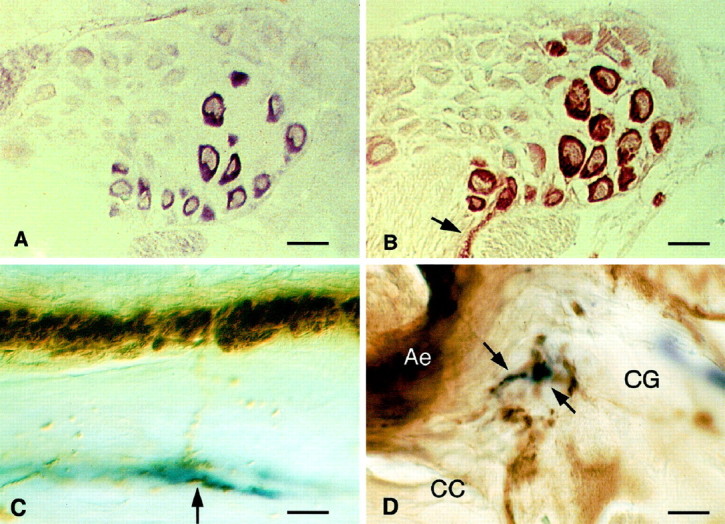
Localization of GRL106 and LyCEP in theLymnaea CNS. A, GRL106 expression revealed by in situ hybridization of a section of theLymnaea CNS with a GRL106-specific probe. Within the cerebral ganglia, one of which is shown here, cells located at the position of CDC neurons hybridize with the GRL106 probe.B, Identification of CDCs by immunohistochemical staining of a section consecutive to the one in A with an antibody against ELH. The cells hybridizing with the GRL106 probe inA are clearly identified as CDCs by their strong reaction with the ELH antibody; in the commissure, immunopositive axon bundles and endings of the CDCs can be seen (arrow). Scale bars: A, B, 80 μm.C, Immunohistochemical double staining of theLymnaea CNS with an anti-ACEP-1 antibody (blue reaction product) and an anti-ELH antibody (brown reaction product). Shown is part of the cerebral commissure, where ELH-positive fibers seem to run to and make contact with fibers that react with the ACEP-1 antibody (arrow). Scale bar, 10 μm. D, In the neuropil of the cerebral ganglion (CG), axons that are immunoreactive to the ACEP-1 antibody and closely opposing axons containing ELH (arrows), which may indicate a site of contact between the LyCEP-expressing neurons and the CDCs. CC, Cerebral commissure containing the axon endings of the CDCs (Ae). Scale bar, 5 μm.