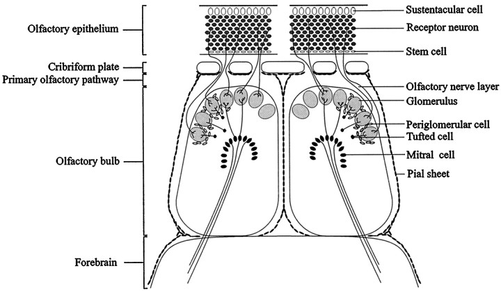
Fig. 1.
Illustration of the anatomical relationships in the primary olfactory system. The cell bodies of olfactory receptor neurons are located in the olfactory epithelium of the nasal cavity and project their axons through the cribriform plate into the olfactory bulb glomeruli, where they terminate on the processes of second-order olfactory neurons: the mitral, tufted, and periglomerular cells. Throughout life, olfactory receptor neurons are replaced continuously from a population of stem cells located in the basal region of the epithelium. As a consequence, newly formed olfactory axons are constantly being extended toward their targets in the main olfactory bulb. To gain further insight into the molecular mechanisms underlying axonal regeneration in the primary olfactory pathway, the expression of the chemorepellent semaphorin III, its receptor neuropilin-1, and CRMP-2 were investigated in the primary olfactory pathway after two lesioning procedures: unilateral olfactory bulbectomy and unilateral transection of the primary olfactory nerve. Injury to the primary olfactory nerve results in degeneration and subsequent replacement of olfactory receptor neurons. After transection of the primary olfactory nerve between the cribriform plate and the olfactory bulb, newly formed primary olfactory axons regenerate into the CNS, establishing synaptic contacts with their targets in the olfactory bulb. Removal of the olfactory bulb (bulbectomy) also induces neurogenesis. However, in adult rodents, a neural scar prevents the regenerating primary olfactory fibers from reaching undamaged areas, e.g., the frontal pole of the cortex.

