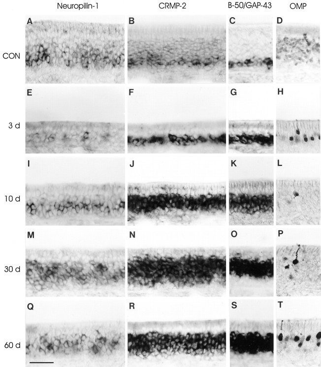
Fig. 2.
Olfactory receptor neurons formed after bulbectomy express neuropilin-1 and CRMP-2 mRNA. Rats subjected to unilateral bulbectomy were allowed to recover for 3 (E–H), 10 (I–L), 30 (M–P), and 60 (Q–T) d. Horizontal cryosections of septal olfactory epithelium from unlesioned animals (CON, 16 weeks of age) and bulbectomized animals were subjected to in situhybridization for neuropilin-1 mRNA (A,E, I, M,Q), CRMP-2 mRNA (B, F,J, N, R), B-50/GAP-43 mRNA (C, G, K,O, S), and immunohistochemistry for OMP protein (D, H, L,P, T). In control epithelium, neuropilin-1 mRNA (A) and CRMP-2 mRNA (B) are expressed in olfactory receptor neurons in the lower region of the olfactory epithelium, corresponding to immature B-50/GAP-43 mRNA-expressing neurons (C) and to a subset of mature OMP-positive neurons directly adjacent to the immature neurons (D). Note that neuropilin-1 and CRMP-2 signals are absent from sustentacular cells and stem cells. As a result of bulbectomy, massive loss of mature OMP-expressing olfactory receptor neurons has occurred. A few OMP-positive neurons remain scattered throughout the ipsilateral epithelium (H,L, P, T). The vast majority of neurons in the bulbectomized epithelium are immature B-50/GAP-43-positive (G, K,O, S). After bulbectomy, neuropilin-1 (E, I, M,Q) and CRMP-2 (F, J,N, R) mRNA expression overlaps with the cohort of immature B-50/GAP-43-expressing neurons. The bulbectomy-induced changes in the mRNA expression for neuropilin-1 and CRMP-2 persist up to at least 60 d after lesion (Q,R). Scale bar, 55 μm.

