
Fig. 4.
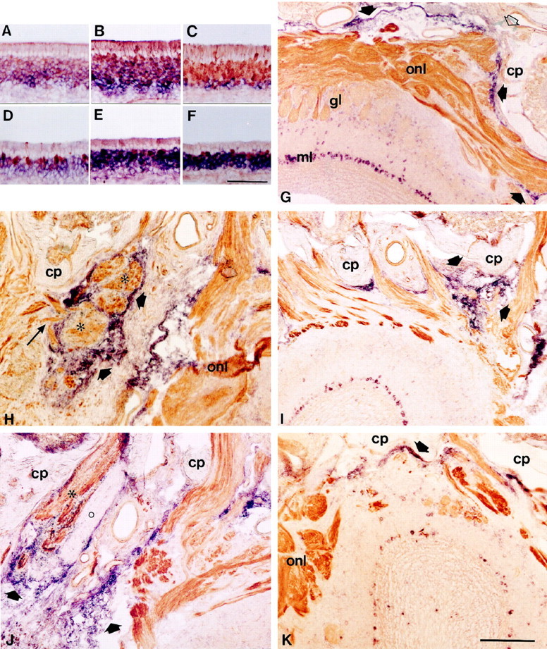
A–F, Double labeling combiningin situ hybridization and immunohistochemistry to examine the expression of neuropilin-1 mRNA and CRMP-2 mRNA in OMP-positive neurons in the intact olfactory epithelium and after bulbectomy. Horizontal sections of olfactory epithelium of unlesioned animals (18 weeks of age) and bulbectomized animals (60 d after lesion) were double stained for neuropilin-1 mRNA (A,D), CRMP-2 mRNA (B, E), or B-50/GAP-43 mRNA (C, F,purple) and OMP protein (brown). Sections probed for neuropilin-1 mRNA were only briefly stained for OMP to allow detection of double-stained profiles. Note that in control epithelium a subset of OMP-expressing mature neurons contains neuropilin-1 and CRMP-2 mRNA (A–C), whereas after bulbectomy, only a small number of neurons express both OMP and neuropilin-1 mRNA or CRMP-2 mRNA (D–F). G–K, Combined in situ hybridization and immunohistochemistry on sections of the olfactory bulb showing the expression of sema III mRNA in the intact situation after bulbectomy or after axotomy and its relation to neuropilin-1-positive olfactory axons. Sections were probed for sema III mRNA (purple) and immunostained for neuropilin-1 protein (brown). Solid arrows point to sema III expression by non-neuronal cells.G, Rostral is to the right. In control olfactory bulb, hybridization signals for sema III are found in non-neuronal cells in the pial sheet and in second-order olfactory neurons. Note that neuropilin-1-positive fibers traverse the cribriform plate through sema III-free spaces (open arrow) and enter the olfactory nerve layer. H–K, Sections of the lesion site at 10 and 60 d after bulbectomy (H,J) or axotomy (I,K). Rostral is to the top, and the contralateral unlesioned side is to the right.H, Non-neuronal cells expressing sema III mRNA surround neuropilin-1-positive olfactory fiber bundles (asterisks) in the lesion cavity at 10 d after bulbectomy. Note that some fibers run through an opening in the rim of sema III-positive cells into a sema III-free region of the scar (long arrow). By 60 d, the bulbar cavity has been invaded by numerous sema III mRNA-containing cells encapsulating neuropilin-1-positive bundles of regenerating olfactory axons (asterisk). In all animals examined, a single encapsulated fiber bundle in the scar consistently lacking immunoreactivity for neuropilin-1 was observed (circle). This neuropilin-1-negative axon bundle might have arisen from olfactory receptor neurons of the vomeronasal epithelium, because these neurons do not express neuropilin-1. At 10 d after axotomy, neuropilin-1-positive fibers are meandering between strings of sema III-positive cells situated between the cribriform plate and the unlesioned olfactory bulb (I). By 60 d, sema III signals in the lesion site have disappeared, and labeling is again confined to the pial sheet (K).cp, Cribriform plate; gl, glomerular layer; onl, olfactory nerve layer. Scale bars: (inF) A–F, 95 μm; (inK) I, J,K, 300 μm; (in K)H, 125 μm.