
Fig. 5.
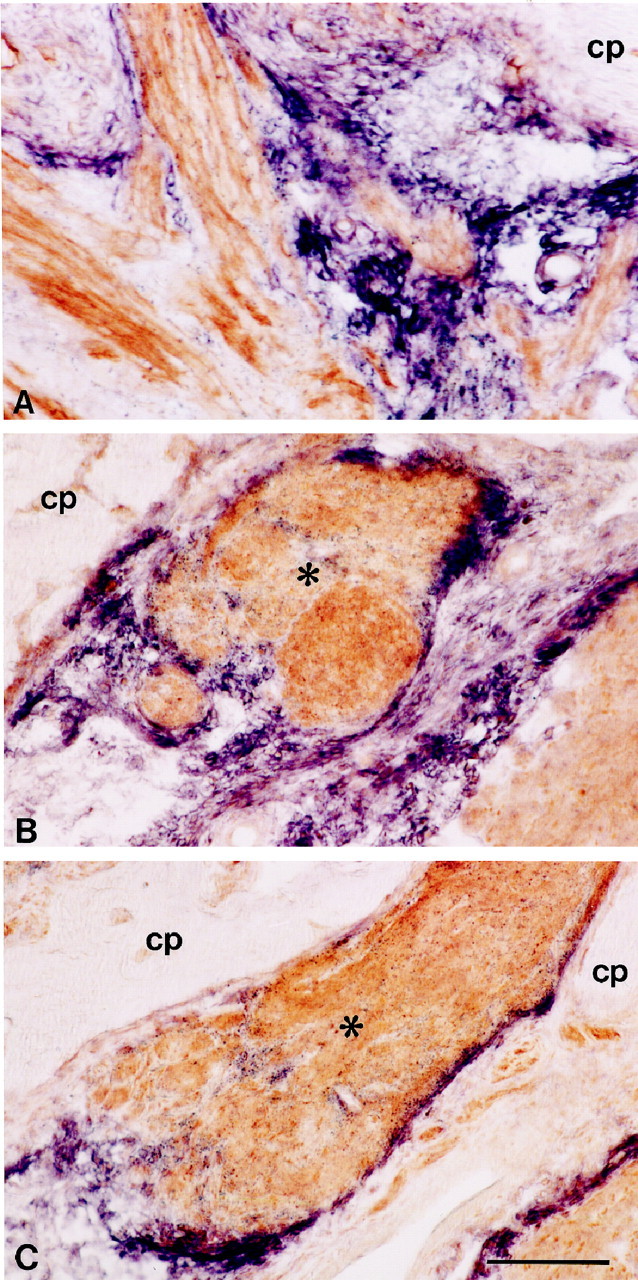
Relationship between regenerating bundles of olfactory axons and sema III mRNA expression in the injured olfactory system. High-power photomicrographs showing horizontal sections of the lesion site at 10 d after transection of the primary olfactory nerve (A) or 60 d after bulbectomy (B, C). Sections were probed for sema III mRNA (purple) and immunolabeled subsequently for neuropilin-1 (A) or B-50/GAP-43 protein (B, C, brown). At 10 d after axotomy, neuropilin-1-positive olfactory axons grow through sema III-free channels lined by strings of sema III mRNA-expressing cells. These cells are continuous with the pial sheet and cover the cribriform plate (A). At 60 d after bulbectomy, bundles of regenerating olfactory axons (asterisks) are tightly encapsulated by sema III-positive cells (B, C). Scale bar, 170 μm.