
Fig. 7.
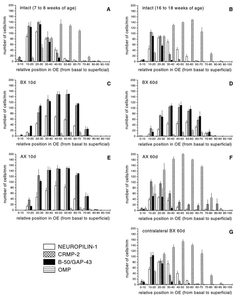
Quantitative assessment of the distribution of neuropilin-1, CRMP-2, B-50/GAP-43, and OMP-positive neurons in the intact and regenerating olfactory epithelium. The number and distribution of neuropilin-1 mRNA, CRMP-2 mRNA, B-50/GAP-43 mRNA, and OMP-positive olfactory receptor neurons were determined at the left side of the septal olfactory epithelium of unlesioned age-matched control animals of 7–8 weeks of age (A), 16–18 weeks of age (B), and at 10 and 60 d after bulbectomy (C, D) or transection of the primary olfactory nerve (E, F). In addition, at 60 d after bulbectomy, the contralateral control side was analyzed (G). For all groups, the number of animals analyzed was three. In young adult animals (7–8 weeks of age), the population of neuropilin-1- and CRMP-2 mRNA-positive neurons overlap almost completely with B-50/GAP-43 mRNA-expressing neurons (A). In fully mature animals (16–18 weeks of age), the cohorts of neuropilin-1- and CRMP-2-expressing neurons overlap with B-50/GAP-43 immature neurons and with OMP-positive mature neurons immediately on top of these immature neurons (B). At 10 d after bulbectomy and axotomy, cohorts of neuropilin-1 mRNA- and CRMP-2 mRNA-expressing neurons display almost complete overlap with B-50/GAP-43 mRNA-containing neurons (C, E). At 60 d after bulbectomy, patterns of expression for neuropilin-1 mRNA and CRMP-2 mRNA resemble those seen at 10 d after lesioning, although the total number of cells has slightly decreased (D). The expression patterns in the contralateral side of the epithelium (G) are indistinguishable from those of unlesioned control animals (B). In contrast to bulbectomy, at 60 d after axotomy, expression patterns strongly resemble control (F). BX, Bulbectomy; AX, axotomy.