
Fig. 3.
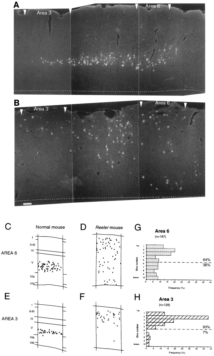
Low-power montage of microphotographs showing the distribution of corticospinal neurons in adult normal (A) and reeler(B) in cortical areas 3, 4, and 6. Area 4 is located between area 3 (to the left) and area 6 (to theright). Microphotographs from rostrocaudal level shown in Figure 1C. The plots shown in C–Fillustrate more closely the differences in radial distribution of corticospinal neurons in area 6 in normal (C) andreeler (D) and in area 3 in normal (E) and reeler(F). Histograms of the radial distribution of corticospinal neurons taken from four to five sections in two to three adult cortices are shown for reeler in area 6 (G) and area 3 (H). To construct these histograms the cortex has been divided into 10; bin 1 is superficial, bin 10 is deep. Scale bar (shown in Bfor A, B): 150 μm.