
Fig. 1.
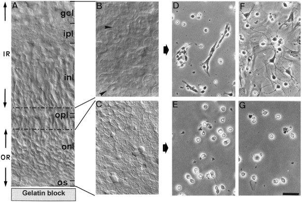
Photographs showing the successive steps of PR culture preparation. Rat retinas (5 d old) (a transverse section showing the different cell and fiber layers is shown inA) were flat-mounted onto the gelatin block with the ganglion cell layer (gcl) uppermost and sectioned along a horizontal plane using a vibratome. An initial cut of 150 μm permitted isolation of the inner retina (IR), composed of the gcl, inner plexiform layer (ipl), and the majority of the inner nuclear layer (inl). A second cut of 30–40 μm containing the remaining inl, outer plexiform layer (opl), and a fraction of the outer nuclear layer (onl) was eliminated (corresponding to the area delimited by dotted lines in A) to ensure purity of the final fraction. A final cut of 200 μm containing only the outer retina (OR) composed of the onl and PR outer segments (os) was then made. B andC demonstrate the microscopical aspect of the IR and OR horizontal slices, respectively. In IR, neuronal cell bodies of different sizes are visible (B, arrows). OR contains only regularly sized PR (C). Cell cultures were obtained after enzymatic digestion and cell dissociation of IR and OR (D–G). Cultures are shown after 1 d (D, E) and 5 d (F, G)in vitro. IR-derived cultures contained multipolar neurons and glial cells (D), the latter of which proliferated to form a monolayer after 5 d in vitro(F). On this glial monolayer, neurons extended long neuritic processes. In contrast, OR-derived cultures were composed of small round cells isolated or present in small groups, sometimes exhibiting small thin neurites (E). After 5 d in vitro, the number of PRs had decreased, but there was no glial proliferation (G). Scale bars:A, 20 μm; B, C, 12.5 μm;D–G, 30 μm.