
Fig. 2.
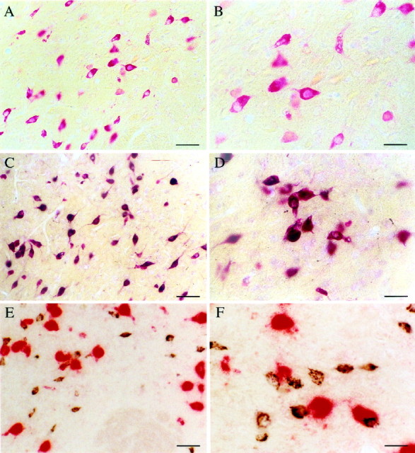
Photomicrographs illustrating hcrt neurons in the perifornical nucleus of the hypothalamus. A, Neurons containing mRNA for hcrt visualized with a homogeneousred coloration of the cytoplasm with an in situ hybridization technique using Fast red as a substrate for alkaline phosphatase. B, Enlargement ofA. C, Photomicrograph showing that all neurons that stained red after in situhybridization (recognizing hcrt mRNA using Fast red) are labeledblack by immunohistochemistry (recognizing the protein with the antiserum #2050) for hcrt. This result indicates that the antiserum #2050 is specific for hcrt. D, High magnification of double-labeled cells after in situhybridization (red) and immunohistochemistry (black). E, Photomicrograph of neurons containing mRNA for the melanin-concentrating hormone (labeled inred by in situ hybridization using Vector red as substrate) and of hcrt neurons (labeled in blackby immunohistochemistry using DAB with nickel as the substrate). Note that no double-labeled cells are present, indicating that MCH and hcrt are found in two distinct populations of neurons. F, High magnification of the MCH (red) and hcrt (black) neurons in the perifornical nucleus. Scale bars:A, C, E, 65 μm;B, D, F, 36 μm.