
Fig. 4.
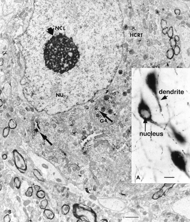
A, Large cells in the lateral hypothalamus are immunoreactive for hypocretin. Staining was found in the cytoplasm and dendrites (short arrow) but not in the nucleus (long arrow). Scale bar, 15 μm.B, Electron microscopic examination of immunoreactive neurons showed punctate staining in the cytoplasm, often associated with dense core granules and parts of the Golgi apparatus (thin arrow). Random organelles near dense core granules sometimes showed peroxidase label, probably because of diffusion during the process of immunocytochemistry. GA, Golgi apparatus; HCRT, hypocretin; NCL, nucleole (thick arrow); NU, nucleus.