
Fig. 7.
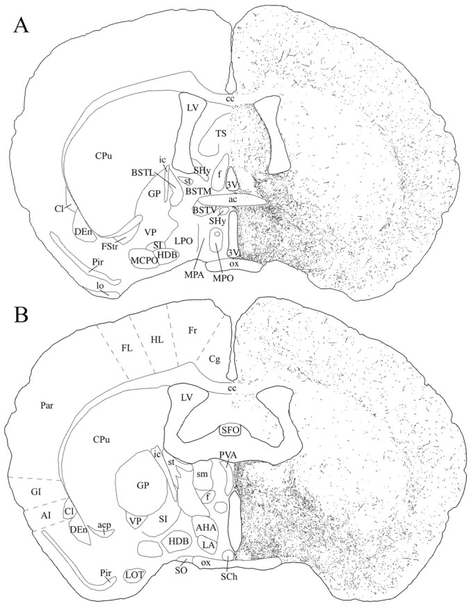
Schematic drawings of 20 μm rostrocaudal coronal sections illustrating the distribution and relative density of hcrt fibers at the level of the preoptic area after immunohistochemistry for hcrt using antibody #2050. 3V, 3rd ventricle;ac, anterior commissure; acp, anterior commissure, posterior part; AHA, anterior hypothalamic area, anterior part; AI, agranular insular cortex;BSTL, bed nucleus of the stria terminalis, lateral division; BSTM, bed nucleus of the stria terminalis, medial division; BSTV, bed nucleus of the stria terminalis, ventral division; cc, corpus callosum;Cg, cingulate cortex; Cl, claustrum; CPu, caudate putamen; DEn, dorsal endopiriform nucleus;f, fornix; FL, forelimb area of the cortex; Fr, frontal cortex; FStr, fundus striati; GI, granular insular cortex; GP, globus pallidus; HDB, nucleus of the horizontal limb of the diagonal band; HL, hindlimb area of the cortex;ic, internal capsule; LA, lateroanterior hypothalamic nucleus; lo, lateral olfactory tract;LOT, nucleus of the lateral olfactory tract;LPO, lateral preoptic area; LV, lateral ventricle; MCPO, magnocellular preoptic nucleus;MPA, medial preoptic area; MPO, medial preoptic nucleus; ox, optic chiasm; Par, parietal cortex; Pir, piriform cortex;PVA, paraventricular thalamic nucleus, anterior part;SCh, suprachiasmatic nucleus; SFO, subfornical organ; SHy, septohypothalamic nucleus;SI, substantia innominata; sm, stria medullaris of the thalamus; SO, supraoptic nucleus;st, stria terminalis; TS, triangular septal nucleus; VP, ventral pallidum.