
Fig. 8.
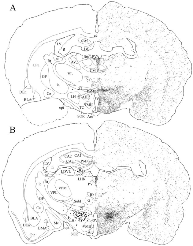
Schematic drawings of 20 μm rostrocaudal coronal sections illustrating the distribution and relative density of hcrt fibers at the level of the tuberal region of the hypothalamus after immunohistochemistry for hcrt using antibody #2050. The position of hcrt cell bodies is indicated as dots in the left hemisphere. 3V, 3rd ventricle; AHP, anterior hypothalamic area, posterior part; Arc, arcuate nucleus; AV, anteroventral thalamic nucleus;BLA, basolateral amygdaloid nucleus, anterior part;BMA, basomedial amygdaloid nucleus, anterior part;CA1–CA3, fields CA1–CA3 of Ammon’s horn;cc, corpus callosum; Ce, central amygdaloid nucleus; CM, central medial thalamic nucleus;CPu, caudate putamen; DEn, dorsal endopiriform nucleus; DG, dentate gyrus;f, fornix; fi, fimbria of the hippocampus; G, gelatinosus thalamic nucleus;GP, globus pallidus; I, intercalated nuclei of the amygdala; ic, internal capsule;LDVL, laterodorsal thalamic nucleus, ventrolateral part;LH, lateral hypothalamic area; LHb, lateral habenular nucleus; LV, lateral ventricle;Me, medial amygdaloid nucleus; mt, mammillothalamic tract; opt, optic tract;PaMP, paraventricular hypothalamic nucleus, medial parvocellular part; Pir, piriform cortex;PoDG, polymorph layer of the dentate gyrus;PV, paraventricular thalamic nucleus;PVA, paraventricular thalamic nucleus, anterior part;Re, reuniens thalamic nucleus; Rh, rhomboid thalamic nucleus; Rt, reticular thalamic nucleus; sm, stria medullaris of the thalamus;SOR, supraoptic nucleus, retrochiasmatic part;st, stria terminalis; SubI, subincertal nucleus; TC, tuber cinereum area; VL, ventrolateral thalamic nucleus; VMH, ventromedial hypothalamic nucleus; VPL, ventral posterolateral thalamic nucleus; VPM, ventral posteromedial thalamic nucleus; ZI, zona incerta.