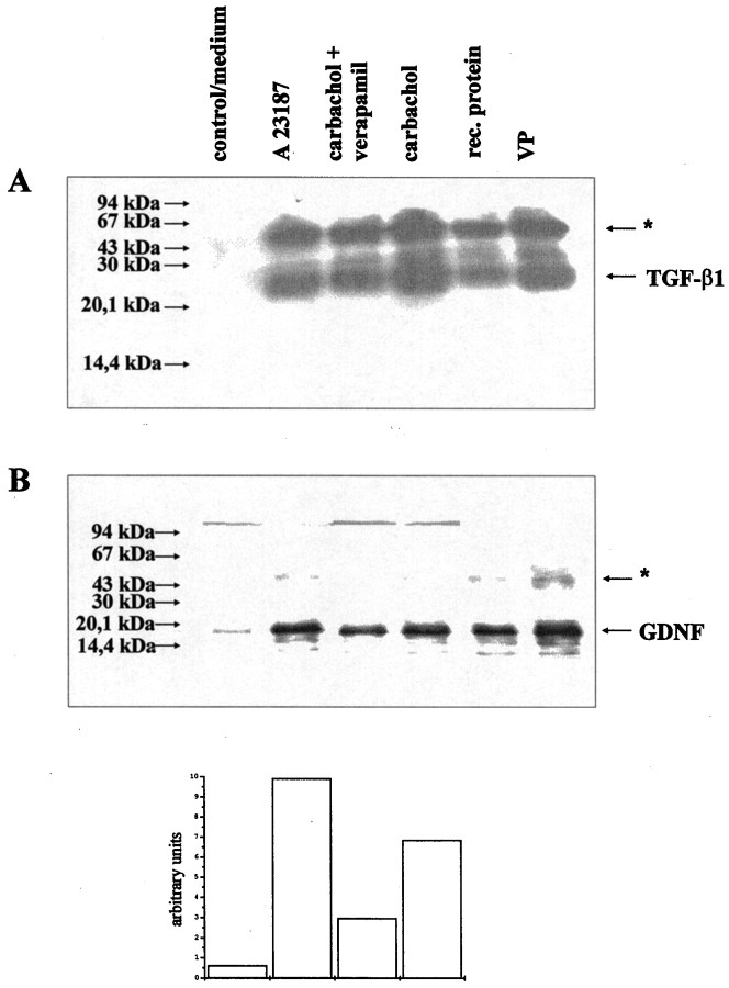
Fig. 10.
GDNF as well as TGF-β1 are released from bovine chromaffin cells after cholinergic stimulation. A, Western blot showing immunoprecipitated TGF-β1 from supernatants of bovine chromaffin cells after a 15 min stimulation with medium only, with a calcium ionophore A23187, carbachol plus verapamil, or carbachol alone. For comparison, rhTGF-β1 and protein extracts from isolated bovine chromaffin granules (VP) were used. Theasterisks indicate the position of the primary antibody used for immunoprecipitation. B, Western blot showing immunoprecipitated GDNF from the same supernatants of bovine chromaffin cells as used for A. For comparison, rhGDNF and VP were used. Bands immunopositive for GDNF were quantified densitometrically and given as arbitrary units.

