
Fig. 6.
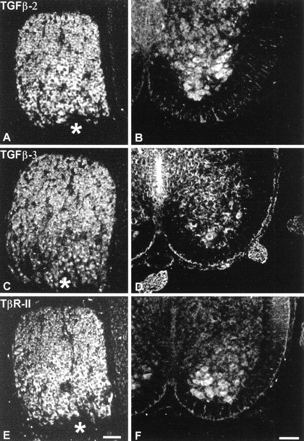
Immunohistochemistry showing localization of TGF-β2 (A, B), TGF-β3 (C, D), and TβR-II (E,F) in chicken DRG (E8; A,C, E) and in rat spinal cord motoneurons (E14; B, D, F). Note strong immunoreactivities in the cell bodies of DRG and motoneurons. Asterisks mark the entrance of dorsal root fibers into the ganglion. Scale bars, 50 μm.