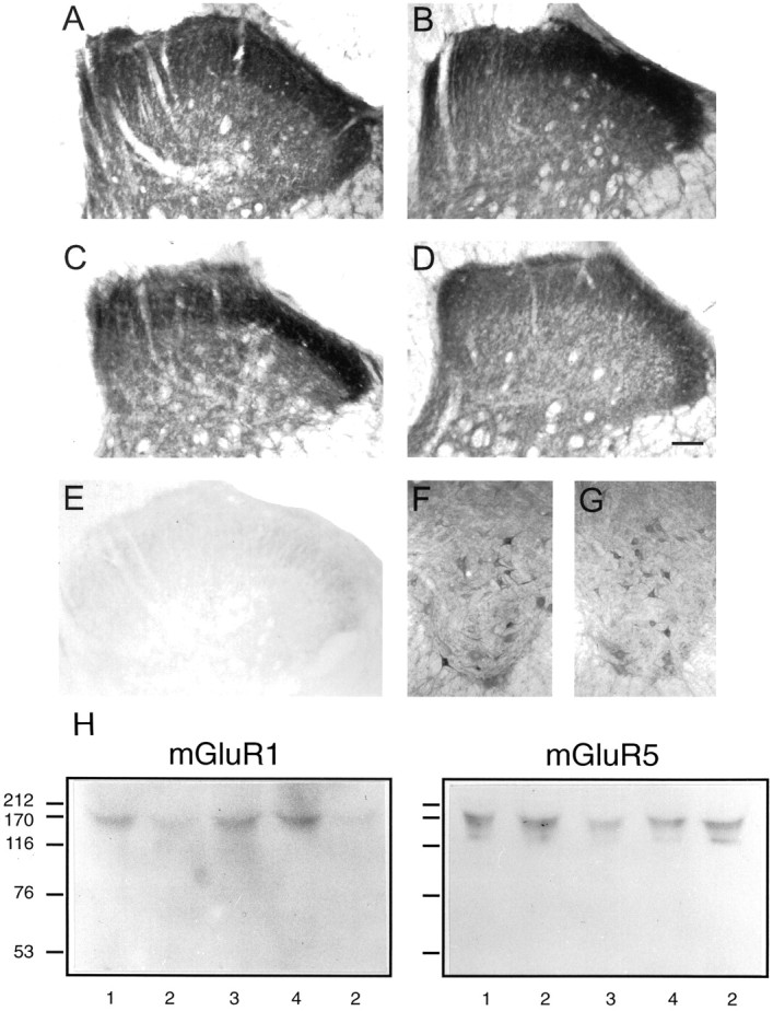
Fig. 3.

Effects of mGluR1 sense, mismatch, and antisense infusion on mGluR1 and mGluR5immunoreactivity in lumbar spinal cord.A–D show typical representations of mGluR1 immunoreactivity in dorsal horn in control (saline), sense, mismatch, and antisense reagent-treated animals.E shows the virtual lack of immunoreactivity in control dorsal horn when the mGluR1 antibody was preabsorbed with membranes from mGluR1-overexpressing cells.F and G show mGluR1immunoreactivity in ventral horn in control and mGluR1antisense-treated animals. These results are typical of at least five animals in each case. Scale bars, 1.0 mm. H shows immunoblots using mGluR1 and mGluR5 antibodies after gel electrophoresis of lysates from spinal cord segments L3–L6 of (1) control, (2) antisense, (3) sense, and (4) mismatch-treated animals. The running positions of the molecular weight markers are shown in kilodaltons. Results are typical of three separate experiments.
