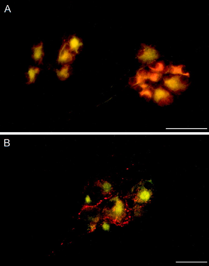
Fig. 1.

Neural elements within the cardiac parasympathetic ganglia exhibit PACAP peptide immunoreactivity. Guinea pig atrial whole-mount preparations were fixed, immunocytochemically labeled for PACAP27 (Cy3, red) and MAP-2 (FITC, green), and examined under fluorescence microscopy. A, PACAP immunoreactivity was localized to a subpopulation of MAP-2-labeled ganglia neurons. B, PACAP peptide staining was prominent in fiber trunks and processes frequently enveloping the principal parasympathetic neurons. Scale bar, 50 μm.
