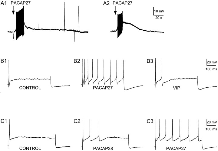
Fig. 8.
PACAP induces depolarization and increases membrane excitability of cardiac ganglia neurons. Local application of PACAP27 (50 μm, 1 sec) depolarized both phasic (A1) and tonic (A2) cardiac neurons. A 500 msec depolarizing current pulse (B, 0.3 nA; C, 0.4 nA) to cardiac neurons typically elicited a single action potential (B1, C1). Superfusion of the same neurons with 100 nm PACAP27 increased membrane excitability under the same depolarizing conditions (B2, C3). In this example, three spikes were elicited after 100 nm PACAP38 application (C2). After a 10 min recovery, the same cell superfused with 100 nm PACAP27 produced seven spikes under an identical depolarizing current pulse (C3). In a comparable experimental paradigm, the changes in neuronal excitability were compared between 100 nm PACAP27 and 100 nm VIP. Nine spikes were elicited after PACAP27 application (B2), whereas only two action potentials were elicited by VIP application in the same neuron after an ∼25 min wash (B3). Additional experiments indicated that 100 nm VIP produced few or no changes in excitability. In all cases, PACAP27 was more effective than the same concentrations of VIP (B3) or PACAP38 (C2). Data are representative of four to five neurons for each treatment paradigm.

